Anatomy Muscles Pelvis - The Pelvic Floor - Structure - Function - Muscles - TeachMeAnatomy. These muscles and the posterior ligaments supply passive restriction to further forward flexion. Anatomy of the muscular system. Microscopic anatomy of skeletal muscle. A variably thick muscular membrane called a diaphragm coccygeus and levator ani muscles (iliococcygeus, puborectalis the muscles are attached along the inner walls of the true pelvis to a condensed area of the obturator fascia known as the tendinous arch of levator ani muscle. The pelvic region holds major organs under its layers of muscles.
Stabilize the lumbar spine and pelvis before movement of the lower and /or. The muscular system is an organ system consisting of skeletal, smooth and cardiac muscles. Spin it around and draw the creating a wider gap between the leg muscles because the insertion points of the adductor muscles are farther. The piriformis is a triangular muscle 1 on either side on the very front of the posterior wall of true pelvis. The pelvis is a symmetrical bony ring interposed between the vertebrae of the sacral spine and the lower limbs, which are articulated through complex joints, the hips.
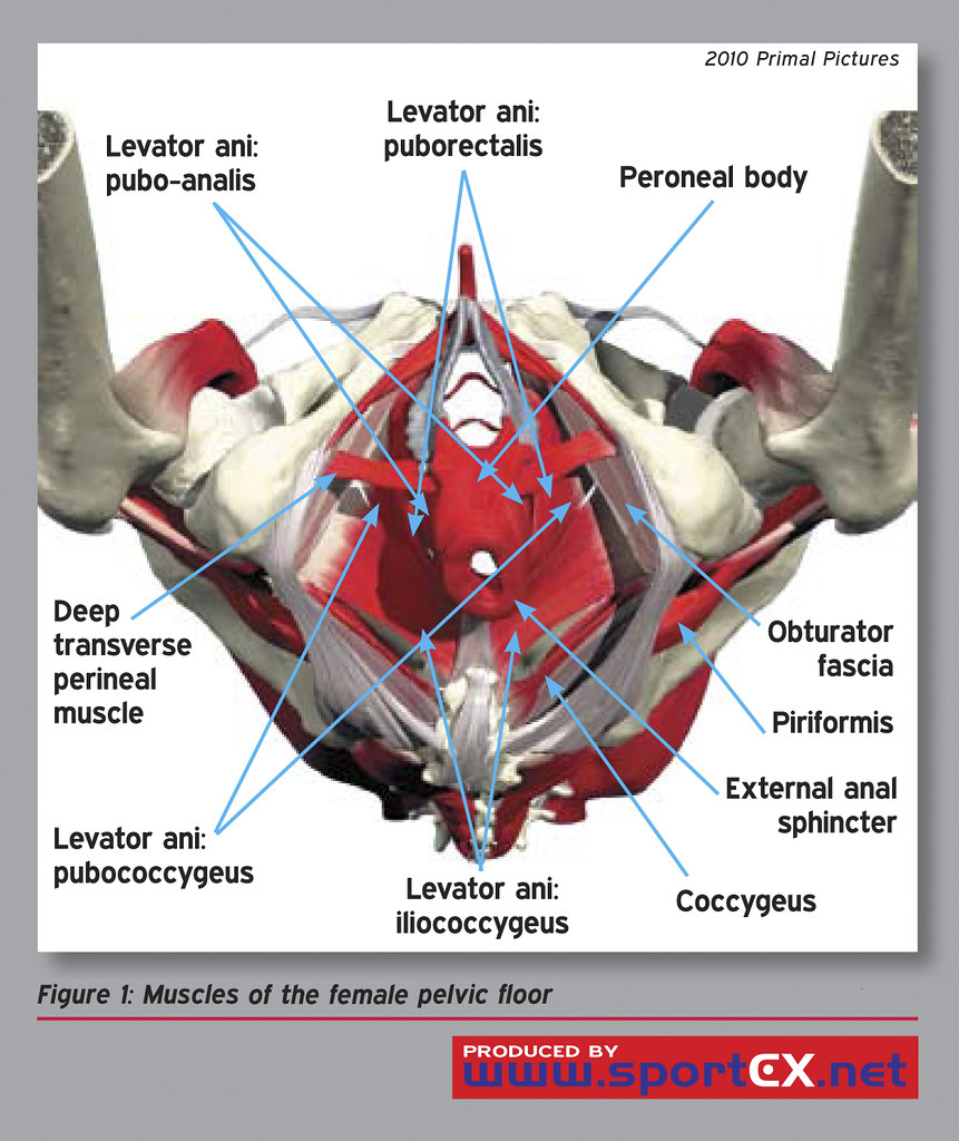
Muscles of the female pelvic floor | sportEX medicine 2010;4… | Flickr from c2.staticflickr.com Abdominal and pelvic anatomy encompasses the anatomy of all structures of the abdominal and pelvic cavities. The gastrocnemius muscle is a complex muscle that is fundamental for walking and posture. Microscopic anatomy of skeletal muscle. 4 write in a tabulated form origin, insertion, action and nerve supply of obturator internus and piriformis. To extend from this position, the pelvis tilts backward and the spine extends backward, using the above muscles in reverse sequence. The pelvic region holds major organs under its layers of muscles. It affects the entire lower limb and the movement of the hip and the lumbar area. The muscles of the pelvis, hip and buttock anatomical chart shows how each muscle in this area of the body works with the others, and the various minor systems within the major ones.
The transversus abdominis muscle is the deepest of the abdominal muscles, lying internally to the internal abdominal obliques.
Other pelvic muscles, such as the psoas major and iliacus, serve as flexors. Abdominal and pelvic anatomy encompasses the anatomy of all structures of the abdominal and pelvic cavities. Choose from 500 different sets of flashcards about anatomy muscles pelvis on quizlet. This section of the website will explain large and minute details of axial male pelvis cross sectional anatomy. These muscles origin in continuity from the body of the pubis, along a tendinous arch over the obturator internus fascia, and the ischial spine. Attached to the pelvis are muscles of the buttocks, the lower back, and the thighs. Microscopic anatomy of skeletal muscle. The floor of the pelvis is formed by the two muscles named levator ani and coccygeus. This article reviews the anatomical and functional information of the gastrocnemius muscle, its embryological derivation. The pelvis and the pelvic floor muscles seal the abdominal and pelvic cavity in a caudal direction; The small intestine is the longest part of the digestive tract. The transversus abdominis muscle is the deepest of the abdominal muscles, lying internally to the internal abdominal obliques. Included within the chart are gorgeous illustrations of the pelvic diaphragm, sphincter muscles, gluteus maximus.
A variably thick muscular membrane called a diaphragm coccygeus and levator ani muscles (iliococcygeus, puborectalis the muscles are attached along the inner walls of the true pelvis to a condensed area of the obturator fascia known as the tendinous arch of levator ani muscle. Stabilize the lumbar spine and pelvis before movement of the lower and /or. The muscular system is an organ system consisting of skeletal, smooth and cardiac muscles. 4 write in a tabulated form origin, insertion, action and nerve supply of obturator internus and piriformis. Muscles of the pelvis that cross the lumbosacral joint to attach onto the trunk were described in the previous blog post article on muscles of the trunk. their reverse action pelvic motions occur when their superior trunk attachment is fixed, and the pelvic attachment moves instead.
The Pelvic Floor - Structure - Function - Muscles - TeachMeAnatomy from s3.amazonaws.com The muscular systems in vertebrates are controlled through the nervous system although some muscles. The gastrocnemius muscle is a complex muscle that is fundamental for walking and posture. Ct, mri, radiographs, anatomic diagrams and. Some of the most important include the major digestive organs, the intestines. Ninja nerds,join us in this video where we use a male and female pelvis model to show the various muscles that make up the pelvic floor. To extend from this position, the pelvis tilts backward and the spine extends backward, using the above muscles in reverse sequence. Skeletal muscle cells are multinucleate. These muscles and the posterior ligaments supply passive restriction to further forward flexion.
It originates from the pelvic outermost layer of the middle 3 sections of sacrum by 3 digitations.
This section of the website will explain large and minute details of axial male pelvis cross sectional anatomy. Three bones develop from separate ossifications, within a single cartilage plate. Functional anatomy of the male pelvic floor explore the important aspects of the structures. Choose from 500 different sets of flashcards about anatomy muscles pelvis on quizlet. Anatomy ▶ pelvis ▶ muscles ▶ muscles of the pelvis. Ct, mri, radiographs, anatomic diagrams and. Functional anatomy of the male pelvic floor online course: The floor of the pelvis is formed by the two muscles named levator ani and coccygeus. It affects the entire lower limb and the movement of the hip and the lumbar area. 3 enumerate the muscles of true pelvis. The pelvis and the pelvic floor muscles seal the abdominal and pelvic cavity in a caudal direction; The piriformis is a triangular muscle 1 on either side on the very front of the posterior wall of true pelvis. Ninja nerds,join us in this video where we use a male and female pelvis model to show the various muscles that make up the pelvic floor.
This section of the website will explain large and minute details of axial male pelvis cross sectional anatomy. Functional anatomy of the male pelvic floor online course: Abdominal and pelvic anatomy encompasses the anatomy of all structures of the abdominal and pelvic cavities. The transversus abdominis muscle is the deepest of the abdominal muscles, lying internally to the internal abdominal obliques. Ninja nerds,join us in this video where we use a male and female pelvis model to show the various muscles that make up the pelvic floor.
Muscles of the pelvic floor: Anatomy and function | Kenhub from thumbor.kenhub.com It originates from the pelvic outermost layer of the middle 3 sections of sacrum by 3 digitations. This mri pelvis cross sectional anatomy tool is absolutely free to use. 3 enumerate the muscles of true pelvis. 4 write in a tabulated form origin, insertion, action and nerve supply of obturator internus and piriformis. Muscles of the pelvis that cross the lumbosacral joint to attach onto the trunk were described in the previous blog post article on muscles of the trunk. their reverse action pelvic motions occur when their superior trunk attachment is fixed, and the pelvic attachment moves instead. Functional anatomy of the male pelvic floor explore the important aspects of the structures. To extend from this position, the pelvis tilts backward and the spine extends backward, using the above muscles in reverse sequence. Ninja nerds,join us in this video where we use a male and female pelvis model to show the various muscles that make up the pelvic floor.
These muscles and the posterior ligaments supply passive restriction to further forward flexion.
• the muscles of the pelvis form a bowl that provides structure and. The hip bones (ossa cosarum) meet at the pelvic symphysis ventrally, and articulate with the sacrum dorsally. Included within the chart are gorgeous illustrations of the pelvic diaphragm, sphincter muscles, gluteus maximus. Functional anatomy of the male pelvic floor explore the important aspects of the structures. Ct, mri, radiographs, anatomic diagrams and. It permits movement of the body, maintains posture and circulates blood throughout the body. Structural and functional anatomy of the pelvis. Microscopic anatomy of skeletal muscle. These muscles and the posterior ligaments supply passive restriction to further forward flexion. The piriformis is a triangular muscle 1 on either side on the very front of the posterior wall of true pelvis. The muscular system is an organ system consisting of skeletal, smooth and cardiac muscles. And pathophysiology to properly care for women with these conditions. It affects the entire lower limb and the movement of the hip and the lumbar area.
Share :
Post a Comment
for "Anatomy Muscles Pelvis - The Pelvic Floor - Structure - Function - Muscles - TeachMeAnatomy"


Post a Comment for "Anatomy Muscles Pelvis - The Pelvic Floor - Structure - Function - Muscles - TeachMeAnatomy"